Author
Germany, Marburg
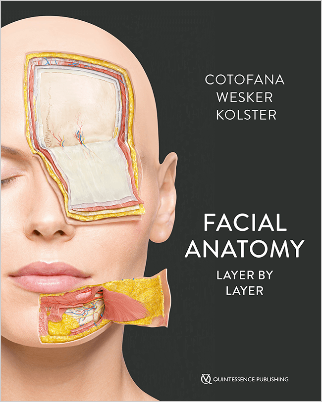
Dr Bernard C. Kolster completed his initial training as a physiotherapist before studying human biology and medicine at the University of Marburg. He subsequently underwent clinical training in physical and rehabilitation medicine as well as other medical disciplines. His particular areas of expertise include the presentation and application of reflex therapy techniques. Dr Kolster is the author and editor of numerous scientific publications and specialist books in the fields of medicine, physiotherapy, naturopathy, and physical and rehabilitation medicine.
Author
Germany, Berlin
Karl H. Wesker is a fine artist and scientific illustrator. For several decades, he has specialized in the clear and didactic visualization of complex anatomical structures. His detailed and aesthetically refined illustrations of human anatomy have appeared in numerous medical publications. Since 1996, he has played a major role in developing the anatomical illustrations for Thieme’s PROMETHEUS Atlas of Anatomy. A particular focus of his work is the clear three-dimensional representation of complex anatomical relationships.
Author
United States of America, Rochester
Professor Sebastian Cotofana is a globally recognized expert in clinical anatomy, whose research has substantially contributed to the detailed understanding of facial anatomy in the context of esthetic medicine. His work emphasizes the anatomical basis of both surgical and minimally invasive procedures, aiming to enhance safety and efficacy in clinical practice. He is the developer of the Cotofana Dissection Method, a systematic, layer-by-layer anatomical approach designed to optimize educational dissection and procedural planning.